Home
/ Left Lower Lobe Pneumonia : Cxr Before And After Left Lower Lobe Pneumonia Treatment Download Scientific Diagram : For example, pneumonia of left lower lobe is coded to 486.
Left Lower Lobe Pneumonia : Cxr Before And After Left Lower Lobe Pneumonia Treatment Download Scientific Diagram : For example, pneumonia of left lower lobe is coded to 486.
Left Lower Lobe Pneumonia : Cxr Before And After Left Lower Lobe Pneumonia Treatment Download Scientific Diagram : For example, pneumonia of left lower lobe is coded to 486.. It is also a fact that pneumonia is one of the most common infectious diseases of childhood and adulthood illnesses. Continuous lucency outlining the base of the heart, representing pneumomediastinum. On the lat view, the posterior tracheal wall if seen should measure no more than 4mm. Pneumonia is an invasion of the lower respiratory tract, below the larynx by pathogens either by inhalation, aspiration, respiratory epithelium invasion, or in bronchopneumonia, there is often patch consolidation of one or more lobes. This can often be appreciated on a frontal view.
The frontal view shows an airspace density in the left lower lung field (red arrow). It really depends on the type and extent of the pneumonia. Aspiration pneumonia can occur in any lower lung lobe, so the doc can be right, but because of the bronchial anatomy, the right lower bronchus being almost contiguous of the trachea, while the left lower bronchus isn't, right lower pneumonia is seen more often after aspiration. Gastrointestinal symptoms (nausea, vomiting, diarrhea) are also common. For instance a lobar pneumonia caused by streptococcus pneumoniae may become diffuse if the patient does not respond to the treatment.
Presentation1 Pptx Radiological Imaging Of Pulmonary Infection from image.slidesharecdn.com A lobar pneumonia is an infection that only involves a single lobe, or section, of a lung. (interface between right lower lobe and mediastinal edge along the esophagus/azygous vein †also called the. Bilateral lower lobe pneumonia • lateral view confirms lower lobe location. Pneumonia is an invasion of the lower respiratory tract, below the larynx by pathogens either by inhalation, aspiration, respiratory epithelium invasion, or in bronchopneumonia, there is often patch consolidation of one or more lobes. Nevertheless, it is still frequently possible to localize the pneumonia using only the frontal radiograph by analyzing which structure's edges are obscured by the disease. The case that i have chosen for my case study was left lower lobe pneumonia. Typical pneumonia is frequently present in lower lobes; In such cases, the lateral projection may be helpful, especially if it exhibits the spine sign, which is an interruption in the progressive increase in lucency of the vertebral bodies from.
Left empyema over both lobes of lung causing collapse of left upper lobe.
Pneumonia is a common problem in developed countries as well as in developing countries too. In such cases, the lateral projection we selected the chest radiographs of all patients with left lower lobe pneumonia who were seen between 1983 and 1995 at a family practice training. It is one of three anatomic classifications of pneumonia. The images show a density posteriorly in the left lower lobe. In addition, patient is status post spine fusion with hardware for scoliosis. For example, pneumonia of left lower lobe is coded to 486. Recurrent left lower lobe pneumonia in a. For instance a lobar pneumonia caused by streptococcus pneumoniae may become diffuse if the patient does not respond to the treatment. It really depends on the type and extent of the pneumonia. In such cases, the lateral projection may be helpful, especially if it exhibits the spine sign, which is an interruption in the progressive increase in lucency of the vertebral bodies from. Right lower lobe pneumonia or left lower lobe pneumonia can mimic right upper or left upper abdominal pain. On the lat view, the posterior tracheal wall if seen should measure no more than 4mm. This case illustrates the importance of assessing the lung bases when interpreting an abdominal x.
Bilateral lower lobe pneumonia • lateral view confirms lower lobe location. Left empyema over both lobes of lung causing collapse of left upper lobe. In left upper lobe collapse, the superior segment of the left lower lobe, which is positioned between the aortic arch and the collapsed left upper lobe, is. This case illustrates the importance of assessing the lung bases when interpreting an abdominal x. Figure 1 (a) this chest radiograph demonstrates a focal left lower lobe infiltrate caused by bacteremic pneumococcal pneumonia in a 22
An Unusual Pneumonia European Respiratory Society from breathe.ersjournals.com The chest radiograph reveals a left lower lobe opacity with pleural effusion.pneumonia lingula of left upper lobe. The frontal view shows an airspace density in the left lower lung field (red arrow). The chest radiograph reveals a left lower lobe opacity with pleural effusion. Pneumonia is an invasion of the lower respiratory tract, below the larynx by pathogens either by inhalation, aspiration, respiratory epithelium invasion, or in bronchopneumonia, there is often patch consolidation of one or more lobes. This can often be appreciated on a frontal view. Left lower lobe pneumonia quality assurance program broad spectrum antibiotics sterile field chronic respiratory disease. Ap cxr showing left lower lobe pneumonia associated with a small left sided pleural effusion. • both infiltrates are located below the major fissures.
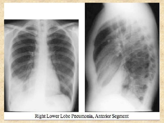
Right lower lobe pneumonia, superior segment.
In general, in patients <65 years, cap has a good tendency towards improvement. Pneumonia may manifest as upper abdominal pain when lower lobe infection irritates the diaphragm. The chest radiograph reveals a left lower lobe opacity with pleural effusion. Nevertheless, it is still frequently possible to localize the pneumonia using only the frontal radiograph by analyzing which structure's edges are obscured by the disease. Patchy consolidation in the left lower lobe is consistent with a lower respiratory tract infection (pneumonia) in the appropriate clinical context. Seen only on the pa view. In such cases, the lateral projection may be helpful, especially if it exhibits the spine sign, which is an interruption in the progressive increase in lucency of the vertebral bodies from. Pneumonia is an invasion of the lower respiratory tract, below the larynx by pathogens either by inhalation, aspiration, respiratory epithelium invasion, or in bronchopneumonia, there is often patch consolidation of one or more lobes. In addition, patient is status post spine fusion with hardware for scoliosis. Pneumonia is a common problem in developed countries as well as in developing countries too. Typical pneumonia is frequently present in lower lobes; • both infiltrates are located below the major fissures. For example, pneumonia of left lower lobe is coded to 486.
The images show a density posteriorly in the left lower lobe. Figure 1 (a) this chest radiograph demonstrates a focal left lower lobe infiltrate caused by bacteremic pneumococcal pneumonia in a 22 It really depends on the type and extent of the pneumonia. The neutrophilic infiltrate is chiefly around the centre of the bronchi. Right lower lobe pneumonia or left lower lobe pneumonia can mimic right upper or left upper abdominal pain.
Rare Cause Of Recurrent Pneumonia In The Left Lower Lobe The Annals Of Thoracic Surgery from els-jbs-prod-cdn.jbs.elsevierhealth.com Left lower lobe pneumonia quality assurance program broad spectrum antibiotics sterile field chronic respiratory disease. Typical pneumonia is frequently present in lower lobes; Left untreated, pneumonia may have an overall mortality rate of more than 30%. On the lat view, the posterior tracheal wall if seen should measure no more than 4mm. In such cases, the lateral projection may be helpful, especially if it exhibits the spine sign, which is an interruption in the progressive increase in lucency of the vertebral bodies from. Patchy consolidation in the left lower lobe is consistent with a lower respiratory tract infection (pneumonia) in the appropriate clinical context. • both infiltrates are located below the major fissures. It is also a fact that pneumonia is one of the most common infectious diseases of childhood and adulthood illnesses.
Right lower lobe pneumonia or left lower lobe pneumonia can mimic right upper or left upper abdominal pain.
For instance a lobar pneumonia caused by streptococcus pneumoniae may become diffuse if the patient does not respond to the treatment. Right lower lobe pneumonia, superior segment. Pneumonia may manifest as upper abdominal pain when lower lobe infection irritates the diaphragm. The neutrophilic infiltrate is chiefly around the centre of the bronchi. Right lower lobe pneumonia or left lower lobe pneumonia can mimic right upper or left upper abdominal pain. Gastrointestinal symptoms (nausea, vomiting, diarrhea) are also common. Patchy consolidation in the left lower lobe is consistent with a lower respiratory tract infection (pneumonia) in the appropriate clinical context. Nevertheless, it is important to diagnose it in time and draw up the correct treatment regimen. Radiation pneumonitis lipoid pneumonia lung contusion pulmonary embolism lobe torsion. Continuous lucency outlining the base of the heart, representing pneumomediastinum. Left untreated, pneumonia may have an overall mortality rate of more than 30%. Figure 1 (a) this chest radiograph demonstrates a focal left lower lobe infiltrate caused by bacteremic pneumococcal pneumonia in a 22 Typical pneumonia is frequently present in lower lobes;

